
In my research and subsequent book "Real-Time Molecular Analysis For Preventing Genetic Diseases," I created a large amount of content and illustrations in reference to emerging technologies to accelerate preventative and precision medicine therapies. I want to share as much of this content as possible. This post will provide an overview of MRI Technologies In Preventative Healthcare. After that, I will cover AI, ML, And DL Technologies, and then Quantum Computing Technologies in an additional post. My primary goal in creating this content was to explain these Interdisciplinary technologies in a manner that would be easily consumable and resonate with a wide audience.
MRI Technologies In Preventative Healthcare
An annual full-body MRI scan is one of the best current methodologies for preventative care. MRI, or Magnetic Resonance Imaging, is a form of non-invasive imaging that uses powerful magnets and radio waves to visualize organs and tissue in the body. An MRI scan can detect potential issues such as heart disease, cancer, and stroke before they become serious. The real benefits come with utilizing Machine Learning from year to year to track changes in the body, monitor for early signs of diseases, and predict potential issues.
For the imaging process, the patient lies down on a table, gliding into an MRI Machine shaped like a cylindrical tube. Using strong magnets, the device produces a magnetic field that aligns the protons in the body's tissues. The protons are then made to produce signals using radio waves, which are subsequently picked up by the machine's receiver and used to create images of the inside of organs. This imaging gives you a very detailed picture of soft tissue parts like the brain, spinal cord, muscles, and organs. Tumors, neurological illnesses, joint and bone issues, and cardiovascular disease are just a few of the conditions that are frequently diagnosed and monitored with this method. MRI uses no ionizing radiation, unlike X-rays and CT scans, making it a safer option for some individuals.
Physical MRI Machine
Generally, MRI Machines are located in hospitals and can be expensive to purchase. A standard physical MRI machine has a powerful magnet, radio frequency coils, and imaging detectors. The most commonly used magnets are 1.5 Tesla or 3 Tesla – with 7 Tesla being the most powerful currently available.

Physical MRI Machine Illustration
MRI Machine Operation Principles
Preparation: Before the scan, the patient must remove any jewelry and change into a gown. They lie down on a table that glides inside the MRI Machine.
Magnetic Field: A strong magnet is needed for the MRI scanner to create a strong magnetic field around the patient. The patient's body's protons align with the magnetic field due to the machine’s magnetic field.
Radio Waves: The protons inside the patient's body produce their own radio waves in response to the radio waves released by the MRI scanner. The machine's receiver picks up these radio waves and uses them to produce an image.
Gradient Coils: Gradient coils cause tiny changes in the magnetic field, which lets the MRI machine make detailed, three-dimensional (3D) pictures of the patient's organs and tissues.
Image Reconstruction: Using sophisticated techniques, a computer reconstructs detailed images of the patient's body from the signals received from the receiver. The photos can be printed out or viewed on a computer screen for additional study.
Primary MRI Components
The major components of the MRI Machine are the casing, outer vacuum shield, outer cold shield, liquid helium vessel, shim coils, magnet, bore, liquid helium cold head, gradient coils, radio frequency coils, inner cold shield, and patient table. The casing of the MRI Machine contains all of the primary components and provides a safe environment from external magnetic fields.
The vacuum shield reduces radiation exposure from the magnet’s radio frequency and gradient coils. Liquid helium is necessary to cool the coils and ensure they don’t overheat. Shim coils are used to adjust the strength of the magnetic field. The magnet itself generates the powerful magnetic field that allows MRI scans to be conducted.
The machine's bore is where the patient enters, and it contains the radio frequency coils that send and receive signals from the body and produce images on a computer screen. The gradient coils create gradients in the magnetic field, allowing images to be taken in different planes. The patient table moves patients into the bore and captures images from various angles.

MRI Primary Components With Callouts Illustration
Magnetic Coils
The magnets are the driving force behind MRI technologies. It all starts with the primary magnetic coil, X magnetic coils, Y magnetic coils, Z magnetic coils, then the radio frequency transmitter and receiver.
The primary magnetic coil is the strongest and most potent of all the coils. It comprises several layers of wire that generate a very strong, stable magnetic field. This field helps to align the hydrogen protons in the body and allows the MRI Machine to capture images.
The X, Y, and Z coils are used to help adjust the magnetic field and create gradients. The radio frequency transmitter and receiver send signals into the body and capture them to create images.

MRI Machine Magnets With Callouts Illustration
High-Level Physics Overview Of MRI Scanning
Magnetization: The hydrogen protons in the patient's body are aligned by the MRI Machine's strong magnetic field. The existence of water and fat molecules causes the body’s abundance of hydrogen protons.
Radiofrequency (RF) Excitation: The hydrogen protons in the MRI Machine absorb radio waves that the machine emits at a precise frequency. The protons become excited as a result, and their magnetic alignment is altered.
Signal Detection: After the RF excitation is stopped, the protons return to their initial magnetic alignment and start to generate their own radio waves. A coil in the MRI Machine detects these impulses and transforms them into electrical signals.
Spatial Encoding: A 2D or 3D image of the patient's body is produced using the signals picked up by the coil. The method used to accomplish this is known as spatial encoding, and it uses gradient magnetic fields that vary throughout the patient's body. The MRI Machine can pinpoint the signal's location within the patient's body by adjusting the strength and timing of the gradient magnetic fields.
Image Reconstruction: A computer reconstructs an image of the patient's body using mathematical methods using signals picked up by the coil. The algorithms allow for producing highly detailed images of internal structures by accounting for the characteristics of various tissues and how they react to the magnetic field and radio waves.
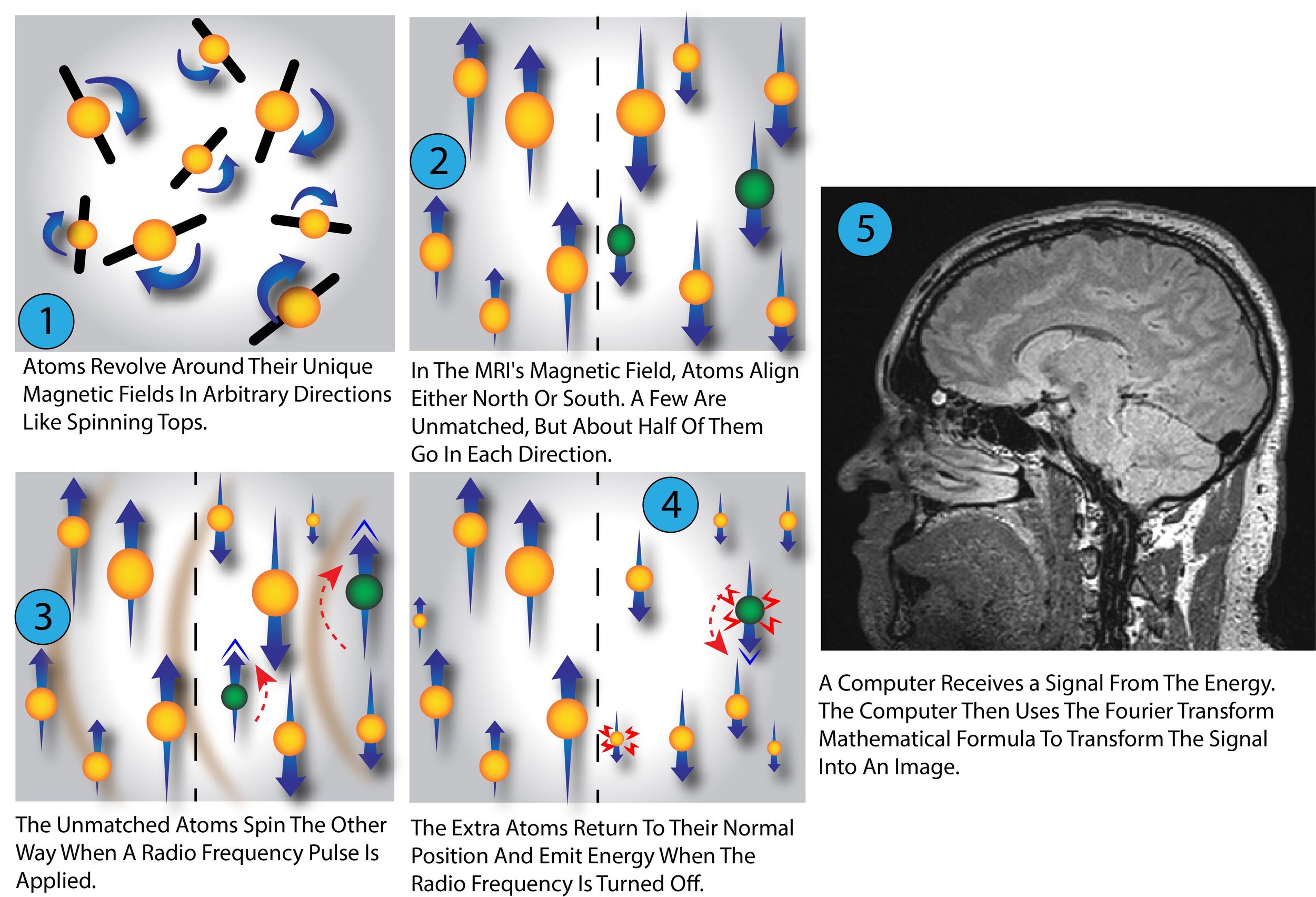
High-Level Physics Overview of MRI Scanning Illustration
MRI Technologies In Preventive And Precision Medicine
MRI technologies are being used in preventive and precision medicine to diagnose diseases earlier and more accurately, improve patient outcomes, reduce costs, and generate personalized treatments. For example, MRI-guided radiation therapy is a procedure that uses an MRI scanner to pinpoint tumors so that doctors can target radiation precisely where it needs to go while avoiding healthy tissue. This has been shown to improve the effectiveness of treatments and reduce side effects.
MRI technologies can also be used to detect early signs of diseases such as cardiovascular disease, stroke, and cancer before they become severe enough to cause noticeable symptoms. MRI scans are often used in conjunction with other imaging modalities such as PET-CT scans, CT scans, and MR spectroscopy to provide more detailed information about a patient's condition.
In addition, MRI technologies have been utilized to develop more personalized treatment plans. By providing better imaging capabilities than other modalities, MRI technologies can detect and examine tumors more granularly, allowing doctors to develop personalized treatment plans for each patient tailored to their specific needs and conditions.
Overall, using MRI technologies in preventative and precision medicine has helped improve patient outcomes while reducing costs. It is an invaluable tool for medical professionals who need accurate information quickly and conveniently to make informed decisions about treatment options for their patients.
Summary
We explored how MRI technologies can be used in preventive and precision medicine. Magnetic Resonance Imaging (MRI) Machines use strong magnetic fields to align hydrogen protons in the patient's body and emit radio waves that the protons absorb, altering their magnetic alignment. The machine then uses a coil to detect the radio waves emitted by the protons as they return to their original magnetic alignment, which is then transformed into electrical signals and analyzed by a computer. This process is known as spatial encoding, which allows for highly detailed images of internal body structures.
MRI technologies have been used in preventive and precision medicine to diagnose diseases earlier and more accurately, improve patient outcomes, reduce costs, and generate personalized treatments. For example, MRI-guided radiation therapy targets cancerous tumors while avoiding healthy tissue. Overall, using MRI technologies has helped improve patient outcomes while reducing costs due to its accuracy and convenience in providing medical professionals with quick access to pertinent information about their patients’ conditions for making informed decisions concerning treatment options.